Radiological Physics and Technology - Cover Gallery
Contents
- Volume 17 (2024) (this opens in a new tab) John Roderik Cameron
- Volume 16 (2023) (this opens in a new tab) Eiichi Tanaka
- Volume 15 (2022) (this opens in a new tab) Russell H. Morgan
- Volume 14 (2021) (this opens in a new tab) Michael Goitein
- Volume 13 (2020) (this opens in a new tab) Michel M. Ter-Pogossian
- Volume 12 (2019) (this opens in a new tab) Charles Edgar Metz
- Volume 11 (2018) (this opens in a new tab) Robert R. Wilson
- Volume 10 (2017) (this opens in a new tab) Louis Harold Gray
- Volume 9 (2016) (this opens in a new tab) Rolf Maximilian Sievert
- Volume 8 (2015) (this opens in a new tab) Antoine Henri Becquerel
- Volume 7 (2014) (this opens in a new tab) Hal Oscar Anger
- Volume 6 (2013) (this opens in a new tab) Kurt Rossmann
- Volume 5 (2012) (this opens in a new tab) Shinji Takahashi
- Volume 4 (2011) (this opens in a new tab) Peter Mansfield and Paul C. Lauterbur
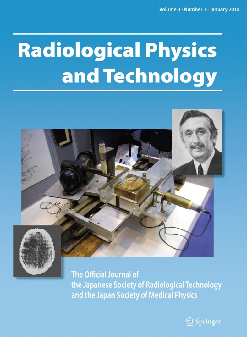
- Volume 3 (2010) (this opens in a new tab) Godfrey N. Hounsfield
- Volume 2 (2009) (this opens in a new tab) Marie Curie
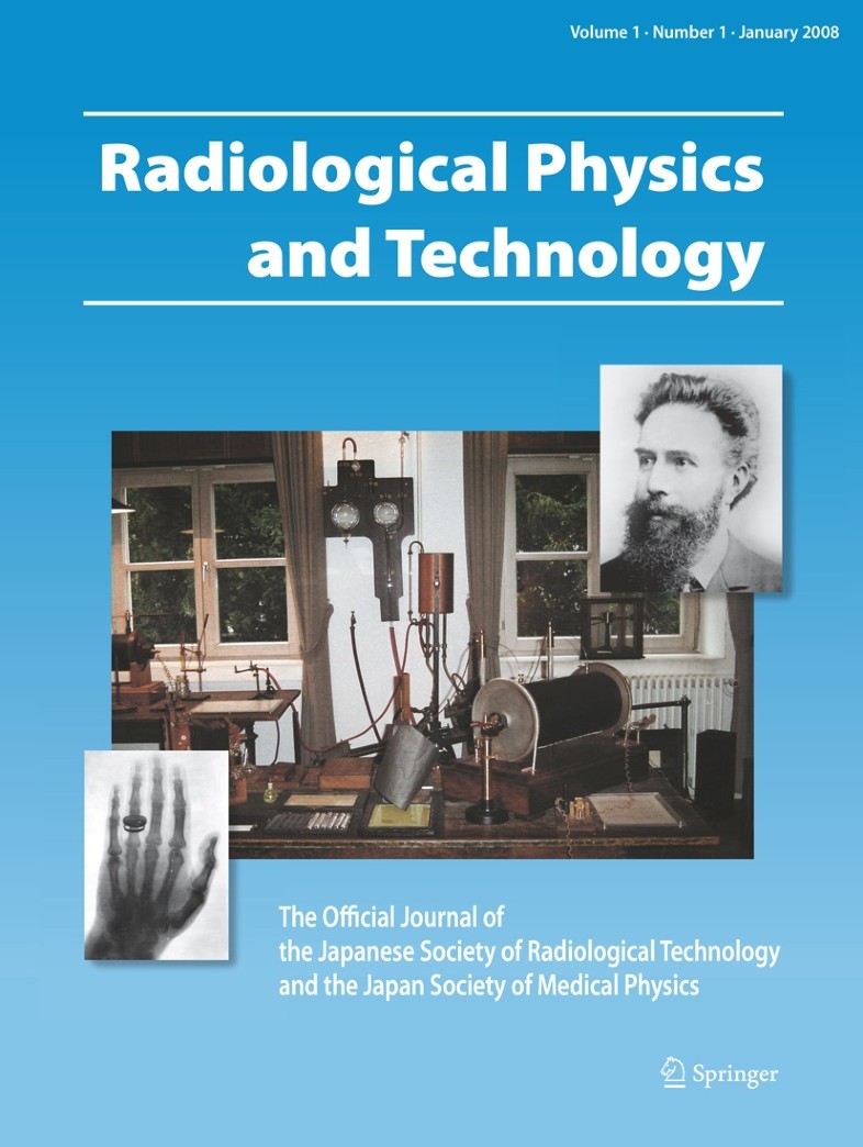
- Volume 1 (2008) (this opens in a new tab) Wilhelm Conrad Röntgen
Volume 17 (2024)
Upper right: John R. Cameron (1922–2005) and Center: Prof. Cameron in his laboratory in 1952, courtesy of the Department of Medical Physics, University of Wisconsin. Lower left: touched-up X-ray showing a dosimeter tube in the rectum near the radioactive source. Values are 24-h dose in cGy. Reprinted from Cameron JR, Daniels F, Kenney G. Radiation dosimeter utilizing the thermoluminescence of lithium fluoride. Science 1961;134:333–4, with permission from AAAS.
John Roderick Cameron (1922–2005): Scientist, teacher, mentor, inventor, and philanthropist extraordinaire (this opens in a new tab) (Editorial by Kwan Hoong Ng & Kunio Doi)
Volume 16 (2023)
Upper right: portrait of Dr. Eiichi Tanaka (1927–2021) by courtesy of Dr. Tanaka’s family. Center (right): Japan’s first positron emission tomography scanner, Positologica-I (© 1979 QST); (left): Toshiba GCA-202 gamma camera with Dr. Tanaka at the age of 45 (© 1972 QST); by courtesy of the National Institutes for Quantum Science and Technology. Lower left: illustrations drawn by Dr. Tanaka, courtesy of Dr. Tanaka’s family.
Eiichi Tanaka, Ph.D. (1927–2021): pioneer of the gamma camera and PET in nuclear medicine physics (this opens in a new tab) (Editorial by Hideo Murayama & Taiga Yamaya)
Volume 15 (2022)
Upper right: Russell H. Morgan, MD (1912-1986), reprinted from Johns Hopkins Radiology with permission. Center and Lower left: Reprinted from Screen intensification systems and their limitations, Ralph E. Sturm and Russell H. Morgan, American Journal of Roentgenology 1949; 62 pp. 617-634, with permission from American Roentgen Ray Society.
Russell H. Morgan, MD (1912–1986): pioneer in image quality assessment and radiological science (this opens in a new tab) (Editorial by Kunio Doi)
Volume 14 (2021)
Upper left: Michael Goitein; Center: Michael Goitein and his colleague evaluating a complex treatment plan, reprinted from International Journal of Radiation Oncology, Biology, Physics, 97, Suit H et al., Michael Goitein, 1939–2016: A Giant of Modern Medical Physics, 654–658, 2017, with permission from Elsevier. Lower left: Beam's-eye view of patient anatomy, reprinted from International Journal of Radiation Oncology, Biology, Physics, 9, Goitein M et al., Multi-dimensional treatment planning: II. Beam’s eye-view, back projection and projection through CT sections, 789–797, 1983, with permission from Elsevier.
Michael Goitein (1939–2016): inventor of three-dimensional planning systems with image-guided beam delivery for radiation therapy (this opens in a new tab) (Editorial by Masahiro Endo & Shinichiro Mori)
Volume 13 (2020)
Upper right: Michel M. Ter-Pogossian (1925–1996). Reproduced with permission from Becker Medical Library, Washington University School of Medicine. Center: Professor Ter-Pogossian with a brain-dedicated scanner PETT-V that he developed. Reproduced with permission from Becker Medical Library, Washington University School of Medicine. Lower left: Cross-section images of a dog obtained with the PETT in the early 1970s. Reproduced from Ter-Pogossian MM et al. A positron-emission transaxial tomograph for nuclear imaging (PETT), Radiology 1975; 114:89–98, with permission from the Radiological Society of North America.
Michel M. Ter-Pogossian (1925–1996): a pioneer of positron emission tomography weighted in fast imaging and Oxygen-15 application (this opens in a new tab) (Editorial by Iwao Kanno, Miwako Takahashi & Taiga Yamaya)
Volume 12 (2019)
Upper right: Charles Edgar Metz, Ph.D. (1942–2012). Center: Figures from the article by Charles E. Metz. Basic principles of ROC analysis. Seminars in Nuclear Medicine. 1978; 8: 283-98; Lower left: Image courtesy of Professor Kunio Doi (Editor in Chief of Radiological Physics and Technology).
Charles Edgar Metz, Ph.D. (1942–2012): pioneer in receiver operating characteristic (ROC) analysis (this opens in a new tab) (Editorial by Junji Shiraishi & Kunio Doi)
Volume 11 (2018)
Upper right: Robert R. Wilson (March 4, 1914 – January 16, 2000), Fermilab, courtesy of AIP Emilio Segrè Visual Archives. Center: Magnets assembled by Albert (Pop) Poperell with his special crew from Bigge Drayage Co. of California. Lower left: The figure of the article by Wilson R. R. on “Radiological Use of Fast Protons.” Radiology 1946; 47:487-491.
Robert R. Wilson (1914–2000): the first scientist to propose particle therapy—use of particle beam for cancer treatment (this opens in a new tab) (Editorial by Masahiro Endo)
Volume 10 (2017)
Upper right: Louis Harold Gray (November 10, 1905 – July 9, 1965). Center: Figures from the article: LH Gray. An Ionization Method for the Absolute Measurement of γ-Ray Energy. Proc. Roy. Soc. A, 156, 578-596, 1936.
Louis Harold Gray (November 10, 1905–July 9, 1965): a pioneer in radiobiology (this opens in a new tab) (Editorial by Masaru Sekiya & Michio Yamasaki)
Volume 9 (2016)
Upper right: Rolf Maximilian Sievert (1896–1966) (taken from the picture collection at the National library of Sweden). Center: Sievert in the physics research laboratory in the Radiumhemmet (licensed under Public domain via Wikimedia Commons) Lower left: External view of the Sievert chamber. Reprinted with permission from Hans Weinberger (Author), Yamasaki Michio (Translator): Life of Sievert — The Father of Radiation Protection, Kokodo Shoten, Niigata, 1994.
Rolf Maximilian Sievert (1896–1966): father of radiation protection (this opens in a new tab) (Editorial by Masaru Sekiya & Michio Yamasaki)
Volume 8 (2015)
Upper left: Antoine Henri Becquerel (1852–1908). Center: Becquerel in the lab. Lower right: An image of uranium crystalline samples recorded on a photosensitive plate, which provided clear evidence in support of invisible radiation emitted by a phosphorescent substance. All photos are licensed under public domain via Wikimedia Commons.
Antoine Henri Becquerel (1852–1908): a scientist who endeavored to discover natural radioactivity (this opens in a new tab) (Editorial by Masaru Sekiya & Michio Yamasaki)
Volume 7 (2014)
Upper left: Hal Oscar Anger (1920–2005), Reprinted by permission of SNMMI from: William G. Myers. Nuclear Medicine Pioneer Citation—1974 Hal Oscar Anger, D.Sc. (Hon.). J Nucl Med. 1974; 15(6): 471–73. Center: The Anger camera invented by Hal O. Anger in 1958. Reprinted from Semin Nucl Med 9(3), Malcolm R. Powell: H. O. Anger and his work at the Donner Laboratory; 164–8, 1979; with permission from Elsevier. Lower right: In vivo 131I image of human thyroid using the first Anger scintillation camera with a 4 in.-diameter NaI(Tl) crystal and 1/4 in.-diameter pinhole. Reprinted with permission from Hal O. Anger: Scintillation Camera. Rev Sci Instrum 29(1):27–33. Copyright 1958, AIP Publishing LLC.
Hal Oscar Anger, D.Sc. (hon.) (1920–2005): a pioneer in nuclear medicine (this opens in a new tab) (Editorial by Hideo Murayama & Tomoyuki Hasegawa)
Volume 6 (2013)
Upper right: Portrait of Kurt Rossmann, Ph.D., 1926–1976. Center: X-ray slit-exposing device for measurements of the LSF and MTF of screen-film systems. Lower left: Radiographs of a needle and plastic beads, obtained with use of two different screen-film systems (reprinted with permission from the American Journal of Roentgenology). All photos appear in the Editorial by Kunio Doi (pp. 1–4).
Kurt Rossmann, Ph.D. (1926–1976): pioneer in image quality evaluation and radiologic imaging research (this opens in a new tab) (Editorial by Kunio Doi)
Volume 5 (2012)
Upper right: Shinji Takahashi, M.D. Center: The first universal rotatography device with the patient in the decubitus position. Lower left: Continuous rotatogram of the lower arm which corresponds to the sinogram (right) and reconstructed crosssectional image obtained from the sinogram (left). All the photos are shown in Kunio Doi et al. (2012).
Shinji Takahashi, M.D. (1912–1985): pioneer in early development toward CT and IMRT (this opens in a new tab) (Editorial by Kunio Doi, Kozo Morita, Sadayuki Sakuma & Masaki Takahashi)
Volume 4 (2011)
Upper left: Peter Mansfield. Upper right: Paul C. Lauterbur (photos by Reuters/AFLO). Center: Prototype MR equipment (reprinted by permission from Peter A. Rinck: Magnetic Resonance in Medicine: The Basic Textbook of the European Magnetic Resonance Forum, 5th edition; ABW Publishers, Berlin, 2003). Lower left: The first MR image (reprinted by permission from Macmillan Publishers Ltd.; Nature 1973; 242: 190–191).
Volume 3 (2010)

Upper right: Godfrey N. Hounsfield; photo from Central Press/Getty Images. Center: Prototype CT scanner; photo taken by Philip Cosson, available for re-use under the terms of the GNU Free Documentation License. Lower left: Image of human brain taken in 1972: from Bautz and Kelender, Der Radiologe 2005; 45(4): 350-5.
Volume 2 (2009)

Upper right: Marie Curie. Center: Marie’s laboratory. Lower left: a radiumgraph by Marie of the contents of a purse. ACJC (the Association Curie et Joliot-Curie, Paris, France)
Volume 1 (2008)

Upper right: Wilhelm Conrad Röntgen: provided by Prof. Kunio Doi. Lower left: Hand of von Kölliker, with permission of Deutsches Röntgen-Museum, Remscheid, Germany. Center: Laboratory, with permission of Röntgen-Gedächtnisstätte, Würzburg, Germany.

